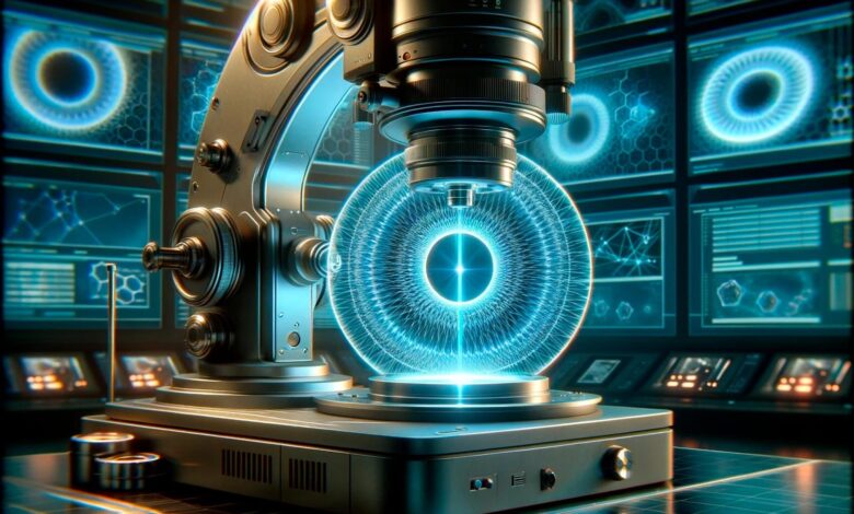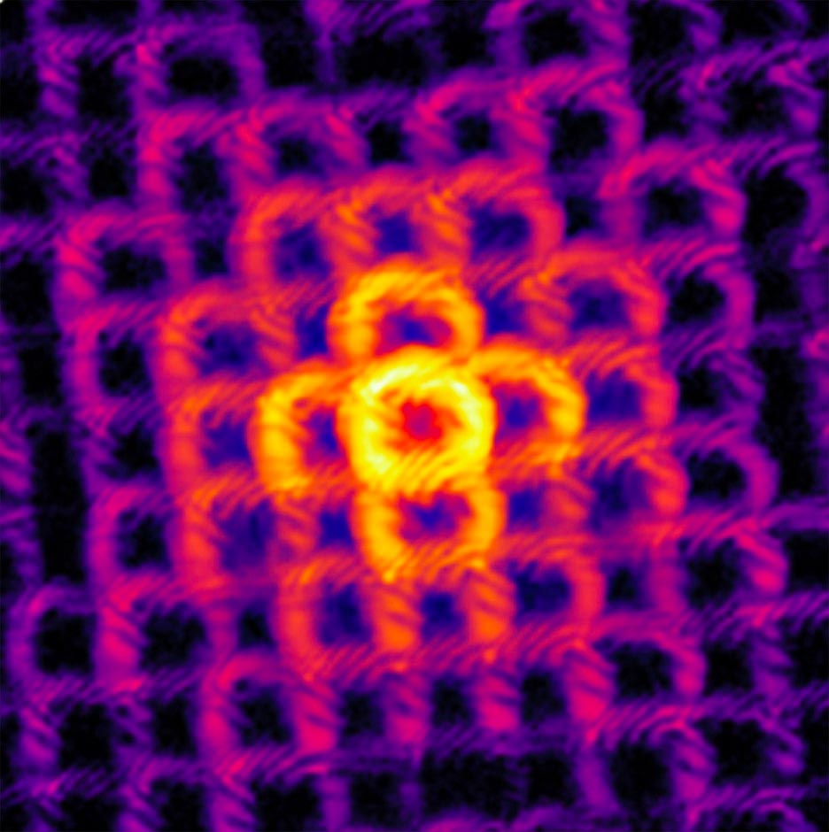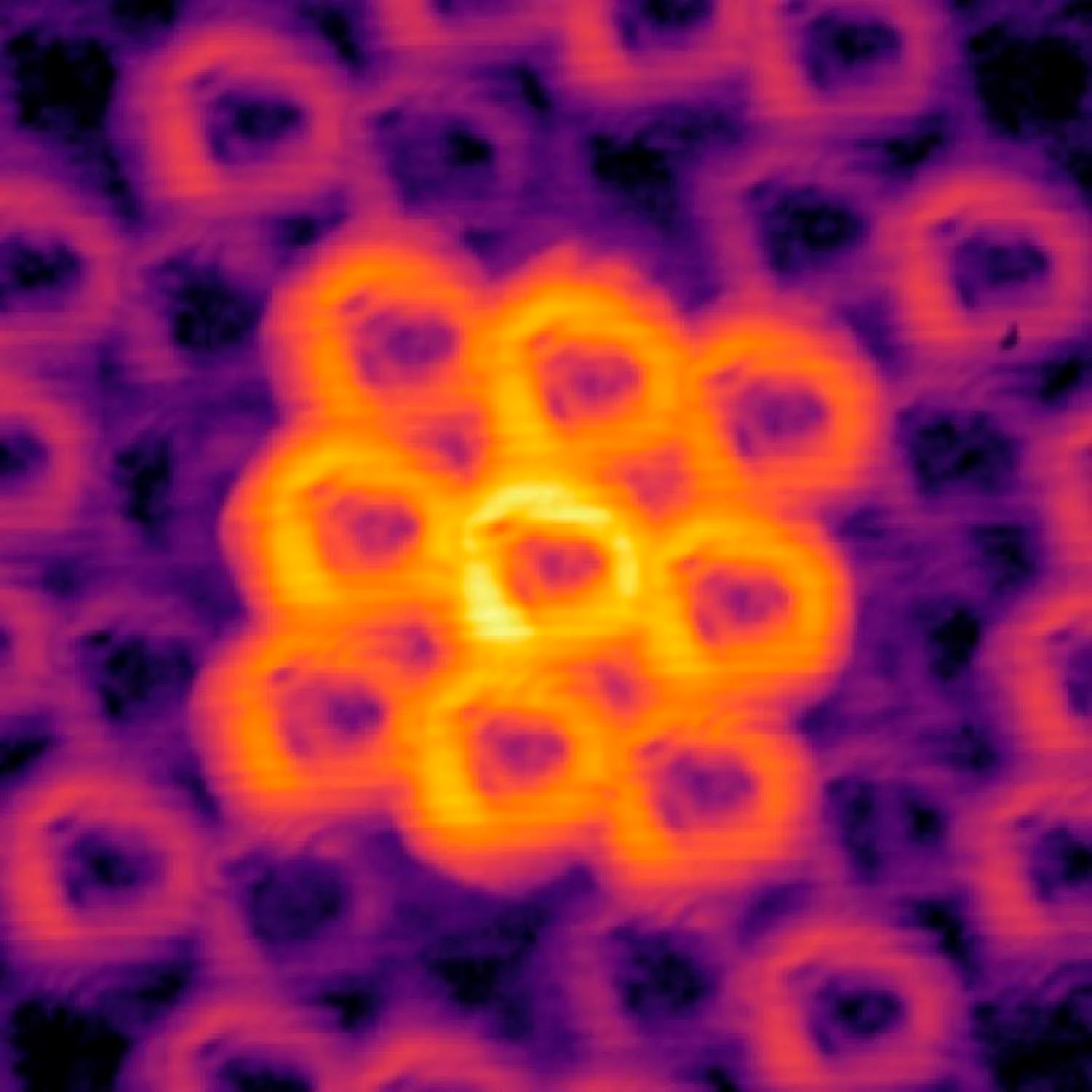
Lead Image: CU Boulder researchers have innovated a new imaging method using doughnut-shaped light beams, advancing the field of ptychography. This technique allows for detailed imaging of tiny, regularly patterned structures like semiconductors, overcoming previous limitations of traditional microscopy. This advancement promises significant improvements in nanoelectronics and biological imaging. (Artist’s concept.) Credit: SciTechDaily.com
In a new study, researchers at CU Boulder have used doughnut-shaped beams of light to take detailed images of objects too tiny to view with traditional microscopes.
Advancements in Nanoelectronics Imaging
The new technique could help scientists improve the inner workings of a range of “nanoelectronics,” including the miniature semiconductors in computer chips. The discovery was highlighted on December 1 in a special issue of Optics & Photonics News called Optics in 2023.
Ptychography: A Lens into the Microscopic World
The research is the latest advance in the field of ptychography, a difficult to pronounce (the “p” is silent) but powerful technique for viewing very small things. Unlike traditional microscopes, ptychography tools don’t directly view small objects. Instead, they shine lasers at a target, then measure how the light scatters away—a bit like the microscopic equivalent of making shadow puppets on a wall.

Overcoming the Ptychography Challenge
So far, the approach has worked remarkably well, with one major exception, said study senior author and Distinguished Professor of physics Margaret Murnane.
“Until recently, it has completely failed for highly periodic samples, or objects with a regularly repeating pattern,” said Murnane, fellow at JILA, a joint research institute of CU Boulder and the National Institute of Standards and Technology (NIST). “It’s a problem because that includes a lot of nanoelectronics.”
She noted that many important technologies like some semiconductors are made up of atoms like silicon or carbon joined together in regular patterns like a small grid or mesh. To date, those structures have proved tricky for scientists to view up close using ptychography.

Breakthrough With Doughnut-Shaped Light
In the new study, however, Murnane and her colleagues came up with a solution. Instead of using traditional lasers in their microscopes, they produced beams of extreme ultraviolet light in the shape of doughnuts.
The team’s novel approach can collect accurate images of tiny and delicate structures that are roughly 10 to 100 nanometers in size, or many times smaller than a millionth of an inch. In the future, the researchers expect to zoom in to view even smaller structures. The doughnut, or optical angular momentum, beams also won’t harm tiny electronics in the process—as some existing imaging tools, like electron microscopes, sometimes can.
“In the future, this method could be used to inspect the polymers used to make and print semiconductors for defects, without damaging those structures in the process,” Murnane said.
Bin Wang and Nathan Brooks, who earned their doctoral degrees from JILA in 2023, were first authors of the new study.
Pushing the Limits of Microscopes
The research, Murnane said, pushes the fundamental limits of microscopes: Because of the physics of light, imaging tools using lenses can only see the world down to a resolution of about 200 nanometers—which isn’t accurate enough to capture many of the viruses, for example, that infect humans. Scientists can freeze and kill viruses to view them with powerful cryo-electron microscopes, but can’t yet capture these pathogens in action and in real time.
Ptychography, which was pioneered in the mid-2000s, could help researchers push past that limit.
The Mechanics of Ptychography
To understand how, go back to those shadow puppets. Imagine that scientists want to collect a ptychographic image of a very small structure, perhaps letters spelling out “CU.” To do that, they first zap a laser beam at the letters, scanning them multiple times. When the light hits the “C” and the “U” (in this case, the puppets), the beam will break apart and scatter, producing a complex pattern (the shadows). Employing sensitive detectors, scientists record those patterns, then analyze them with a series of mathematical equations. With enough time, Murnane explained, they recreate the shape of their puppets entirely from the shadows they cast.
“Instead of using a lens to retrieve the image, we use algorithms,” Murnane said.
She and her colleagues have previously used such an approach to view submicroscopic shapes like letters or stars.
But the approach won’t work with repeating structures like those silicon or carbon grids. If you shine a regular laser beam on a semiconductor with such regularity, for example, it will often produce a scatter pattern that is incredibly uniform—ptychographic algorithms struggle to make sense of patterns that don’t have much variation in them.
The problem has left physicists scratching their heads for close to a decade.

Doughnut Microscopy
In the new study, however, Murnane and her colleagues decided to try something different. They didn’t make their shadow puppets using regular lasers. Instead, they generated beams of extreme ultraviolet light, then employed a device called a spiral phase plate to twist those beams into the shape of a corkscrew, or vortex. (When such a vortex of light shines on a flat surface, it makes a shape like a doughnut.)
The doughnut beams didn’t have pink glaze or sprinkles, but they did the trick. The team discovered that when these types of beams bounced off repeating structures, they created much more complex shadow puppets than regular lasers.
To test out the new approach, the researchers created a mesh of carbon atoms with a tiny snap in one of the links. The group was able to spot that defect with precision not seen in other ptychographic tools.
“If you tried to image the same thing in a scanning electron microscope, you would damage it even further,” Murnane said.
Advancing Towards Finer Details
Moving forward, her team wants to make their doughnut strategy even more accurate, allowing them to view smaller and even more fragile objects—including, one day, the workings of living, biological cells.
Reference: “High-fidelity ptychographic imaging of highly periodic structures enabled by vortex high harmonic beams” by Michael Tanksalvala, Henry C. Kapteyn, Bin Wang, Peter Johnsen, Yuka Esashi, Iona Binnie, Margaret M. Murnane, Nicholas W. Jenkins and Nathan J. Brooks, 19 September 2023, Optica.
DOI: doi:10.1364/OPTICA.498619
Other co-authors of the new study include Henry Kapteyn, professor of physics and fellow of JILA, and current and former JILA graduate students Peter Johnsen, Nicholas Jenkins, Yuka Esashi, Iona Binnie and Michael Tanksalvala.
