
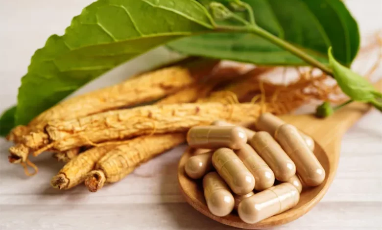
Lead Image: Ginkgo biloba, is commonly known simply as as ginkgo. Because of the way that it is pronounced, gingko is a common alternative spelling.
Ginkgo biloba is an herbal supplement possibly best known for enhancing cognitive performance. However, its benefits and uses are wide-ranging. For example, ginkgo can be taken as an aphrodisiac, as well as a remedy for PMS, headaches, migraines, and even anxiety. It also provides overall protective benefits for your health through a variety of antioxidants.
What is Ginkgo Biloba?
Ginkgo biloba is a medicinal herb that comes from a tree also known as the maidenhair tree. Among the oldest tree species in the world, ginkgo trees are considered “living fossils” because they’ve survived planetary extinction events. Maidenhair trees grow at least about 130 feet tall and can live well over a thousand years.[1]
Ginkgo biloba leaves have been dried and ground into powder in traditional Chinese medicine for thousands of years. The dried ginkgo leaves were also used to make tea. Today, you can find ginkgo biloba pills and ginkgo extract liquid drops in health food stores.
8 Benefits of Ginkgo Biloba
People take ginkgo biloba for a range of purposes. According to what research has uncovered, here are the health benefits ginkgo biloba can provide:
1. Combats ADHD
Ginkgo biloba improves the flow of blood to the brain, causing measurable increases in blood circulation in the visual cortex.[2] In a 6-week study on children with ADHD, ginkgo biloba improved symptoms better than a placebo.[3] While ginkgo supplementation may be helpful for people with difficulty concentrating, it doesn’t necessarily enhance an already optimally-functioning brain.
2. Protects the Aging Brain
Ginkgo shows promise in improving symptoms for dementia patients, which suggests it may also help protect against neurodegeneration in healthy individuals.[4] In a meta-analysis of studies involving ginkgo biloba and Alzheimer’s patients, researchers concluded that 3 to 6 months of ginkgo biloba supplementation can improve cognitive function in patients with Alzheimer’s disease.[5]
3. May Help with Anxiety
In a 4-week study on 107 people with generalized anxiety disorder, 480 mg of ginkgo biloba per day improved symptoms significantly relative to a placebo.[6] Another study suggests that these results come from ginkgo’s ability to modulate gamma-aminobutyric acid (GABA) receptors.[7]
4. Boosts Libido
One of the common causes of suppressed sex drive or low libido is the use of anti-depressants. In a study on men and women taking serotonin reuptake inhibitors (SSRIs) and other types of anti-depressants, ginkgo biloba improved sexual desire, lubrication, and orgasm in 84% of the participants. Among the female participants, the success rate was 91%.[8] Other studies have found that ginkgo biloba effectively boosts libido and sexual function in postmenopausal women.[9]
5. Helps Relieve PMS
Ginkgo biloba has been found in studies to improve symptoms of PMS in women. In one study that looked at 165 women over three menstrual cycles, breast tenderness and pain were reduced by ginkgo biloba, as well as neuropsychological symptoms.[10] Another study that looked at just two consecutive cycles found that 40 mg of ginkgo biloba leaf extract improved symptoms of PMS better than a placebo, including both physical and psychological symptoms.[11]
6 Treats Some Headaches and Migraines
In traditional Chinese medicine, ginkgo was prescribed for treating headaches and migraines, though modern research hasn’t been done to confirm its efficacy. It could work by lowering inflammation and improving cerebral blood flow. However, headaches with other causes may not respond to ginkgo biloba.
7. Supports Heart Health
Many of ginkgo’s benefits come from its ability to boost blood circulation. According to traditional Chinese medicine, ginkgo actually opens channels in the body and allows vital organs like the lungs, liver and brain to be stimulated by “chi.” In people at risk of heart attack and stroke, the blood vessels generally aren’t able to dilate sufficiently for blood to flow.
In a study published in Phytotherapy Research, participants given ginkgo biloba had immediate increases in blood flow and a 12% increase in nitric oxide. Nitric oxide is vital to heart health, as it helps dilate blood vessels.[12] In another study, healthy older adults experienced increased blood flow and blood vessel dilation with ginkgo biloba, suggesting it may help lower the risk of developing heart disease in elderly people.[13]
8. Provides Antioxidant Support
Besides its ability to reduce inflammation and boost blood flow, ginkgo’s powerful antioxidant action is also responsible for many of its health benefits. The antioxidants in ginkgo biloba can potentially improve anxiety, help in the prevention of cancer, as well as protect against neurodegenerative diseases.[14] Antioxidants work by neutralizing free radicals that enter the body from the environment or as a result of normal metabolic functions. By preventing oxidative damage caused by free radicals, antioxidants protect against cellular and DNA damage, in turn, protecting against chronic diseases.
Ginkgo Biloba Dosage and Side Effects
Side effects of ginkgo biloba are rare, except when taken in doses above the recommended limit. The best dose for ginkgo is between 60 and 240 mg for 6 months at a time.[15] However, you should always follow the recommendation on the label of the product you’re using. Potential side effects of ginkgo can include headaches, dizziness, heart palpitations, and gastrointestinal dysfunction.
Can Ginkgo Biloba Benefit Your Health?
Ginkgo biloba is an herb that’s been cultivated as medicine in Asia for millennia. It has many healing properties that help with a wide range of health issues. As an herbal supplement, ginkgo can potentially help ward off diseases. As a natural remedy, you can use it to improve circulation, replenish your sex drive, relieve PMS symptoms or even release a headache.
Keep in mind that even natural substances can potentially cause unwanted side effects. To ensure your safety, it is recommended to consult with a healthcare provider before using herbal products or other supplements, especially if you have existing medical conditions, are taking other medications or supplements, or are pregnant.
References:
- “Health benefits of Gingko biloba” medicalnewstoday.com/articles/263105
- “Examining Brain-Cognition Effects of Ginkgo Biloba Extract: Brain Activation in the Left Temporal and Left Prefrontal Cortex in an Object Working Memory Task” by R. B. Silberstein, A. Pipingas, J. Song, D. A. Camfield, P. J. Nathan and C. Stough, 18 Aug 2011, Evidence-Based Complementary and Alternative Medicine.
DOI: 10.1155/2011/164139 - “Ginkgo biloba in the treatment of attention-deficit/hyperactivity disorder in children and adolescents. A randomized, placebo-controlled, trial” by Fereshteh Shakibaei, Mehrsa Radmanesh, Elham Salari and Behzad Mahaki, 18 April 2015, Complementary Therapies in Clinical Practice.
DOI: 10.1016/j.ctcp.2015.04.001 - “Effects of Ginkgo biloba in dementia: systematic review and meta-analysis” by Stefan Weinmann, Stephanie Roll, Christoph Schwarzbach, Christoph Vauth and Stefan N Willich, 17 March 2010, BMC Geriatrics.
DOI: 10.1186/1471-2318-10-14 - “The Efficacy of Ginkgo biloba on Cognitive Function in Alzheimer Disease” by Barry S. Oken, MD; Daniel M. Storzbach, PhD and Jeffrey A. Kaye, MD, November 1998, JAMA Neurology.
DOI: 10.1001/archneur.55.11.1409 - “Ginkgo biloba special extract EGb 761® in generalized anxiety disorder and adjustment disorder with anxious mood: A randomized, double-blind, placebo-controlled trial” by H. Woelk, K. H. Arnoldt, M. Kieser and R.Hoerr, 30 June 2006, Journal of Psychiatric Research.
DOI: 10.1016/j.jpsychires.2006.05.004 - “GABA-modulating phytomedicines for anxiety: A systematic review of preclinical and clinical evidence” by Karen Savage, Joseph Firth, Con Stough and Jerome Sarris, 23 November 2017, Phytotherapy Research.
DOI: doi.org/10.1002/ptr.5940 - “Ginkgo biloba for antidepressant-induced sexual dysfunction” by Alan J. Cohen and Barbara Bartlik, 14 January 2008, Journal of Sex & Marital Therapy.
DOI: 10.1080/00926239808404927 - “A systematic review of clinical trials on Ginkgo (Ginkgo biloba) effectiveness on sexual function and its safety” by Zahra Niazi Mashhadi, Morvarid Irani, Mahin Kiyani Mask and Clara Methie, July/August 2021, Avicenna Journal of Phytomedicine.
DOI: 10.22038/ajp.2021.17813 - “Value of standardized Ginkgo biloba extract (EGb 761) in the management of congestive symptoms of premenstrual syndrome” by A Tamborini and R Taurelle, July-September 1993, Rev Fr Gynecol Obstet.
PMID: 8235261 - “A Randomized, Placebo-Controlled Trial of Ginkgo biloba L. in Treatment of Premenstrual Syndrome” by Giti Ozgoli, Elham Alsadat Selselei, Faraz Mojab and Hamid Alavi Majd, 13 August 2009, The Journal of Alternative and Complementary Medicine.
DOI: 10.1089/acm.2008.0493 - “Ginkgo biloba extract improves coronary artery circulation in patients with coronary artery disease: contribution of plasma nitric oxide and endothelin-1” by Yu-Zhou Wu, Shu-Qin Li, Xiu-Guang Zu, Jun Du and Feng-Fei Wang, 29 April 2008, Phytotherapy Research.
DOI: 10.1002/ptr.2335 - “Ginkgo biloba extract improves coronary blood flow in healthy elderly adults: Role of endothelium-dependent vasodilation” by Yuzhou Wu, Shuqin Li, Wei Cui, Xiuguang Zu, Jun Du and Fengfei Wang, 6 February 2008, Phytomedicine.
- “Neuroprotective and Antioxidant Effect of Ginkgo biloba Extract Against AD and Other Neurological Disorders” by Sandeep Kumar Singh, Saurabh Srivastav, Rudolph J. Castellani, Germán Plascencia-Villa and George Perry, 2 August 2019, Neurotherapeutics.
DOI: 10.1007/s13311-019-00767-8 - “Ginkgo – Uses, Side Effects, And More” webmd.com/vitamins/ai/ingredientmono-333/ginkgo
Please note that the information contained in this article is for informational purposes only and should not be used as a substitute for professional medical advice, diagnosis, or treatment. If you have any questions or concerns about your health, it is important to speak with a qualified healthcare provider. This article is not intended to diagnose, treat, cure, or prevent any disease. It is not intended to provide medical advice.