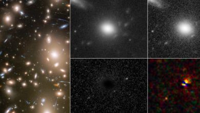
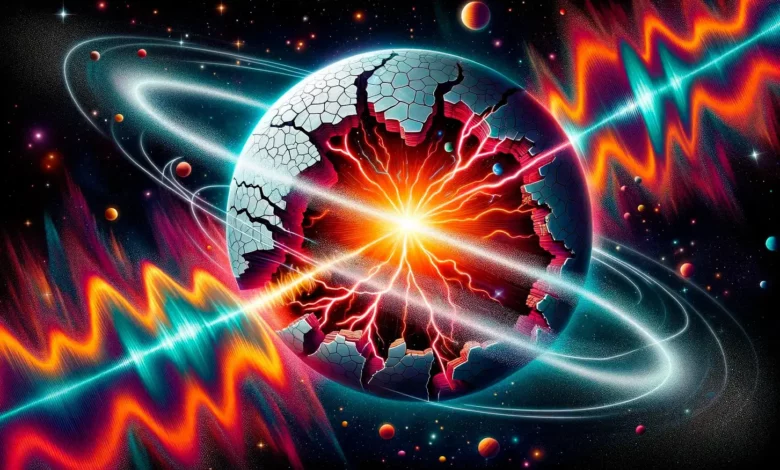
Lead Image: Fast radio bursts (FRBs), intense radio energy bursts, are a perplexing astronomical enigma. The University of Tokyo’s recent research has found similarities between FRBs and earthquakes, suggesting that they might be caused by “starquakes” on neutron stars. These insights could reshape our understanding of earthquakes, high-density matter, and nuclear physics.
Research from the University of Tokyo links fast radio bursts (FRBs) to “starquakes” on neutron stars, offering new insights into earthquakes and nuclear physics.
Fast radio bursts, or FRBs, are an astronomical mystery, with their exact cause and origins still unconfirmed. These intense bursts of radio energy are invisible to the human eye, but show up brightly on radio telescopes. Previous studies have noted broad similarities between the energy distribution of repeat FRBs, and that of earthquakes and solar flares.
However, new research at the University of Tokyo has looked at the time and energy of FRBs and found distinct differences between FRBs and solar flares, but several notable similarities between FRBs and earthquakes. This supports the theory that FRBs are caused by “starquakes” on the surface of neutron stars. This discovery could help us better understand earthquakes, the behavior of high-density matter, and aspects of nuclear physics.
The Enigma of FRBs
The vastness of space holds many mysteries. While some people dream of boldly going where no one has gone before, there is a lot we can learn from the comfort of Earth. Thanks to technological advances, we can explore the surface of Mars, marvel at Saturn’s rings, and pick up mysterious signals from deep space. Fast radio bursts are hugely powerful, bright bursts of energy that are visible on radio waves.
First discovered in 2007, these bursts can travel billions of light years but typically last mere thousandths of a second. It has been estimated that as many as 10,000 FRBs may happen every day if we could observe the whole sky. While the sources of most bursts detected so far appear to emit a one-off event, there are about 50 FRB sources that emit bursts repeatedly.
Theories Behind the Cause of FRBs
The cause of FRBs is unknown, but some ideas have been put forward, including that they might even be alien in origin. However, the current prevailing theory is that at least some FRBs are emitted by neutron stars. These stars form when a supergiant star collapses, going from eight times the mass of our sun (on average) to a superdense core only 20-40 kilometers across. Magnetars are neutron stars with extremely strong magnetic fields, and these have been observed to emit FRBs.
“It was theoretically considered that the surface of a magnetar could be experiencing a starquake, an energy release similar to earthquakes on Earth,” said Professor Tomonori Totani from the Department of Astronomy at the Graduate School of Science. “Recent observational advances have led to the detection of thousands more FRBs, so we took the opportunity to compare the now large statistical data sets available for FRBs with data from earthquakes and solar flares, to explore possible similarities.”
Statistical Analysis and Findings
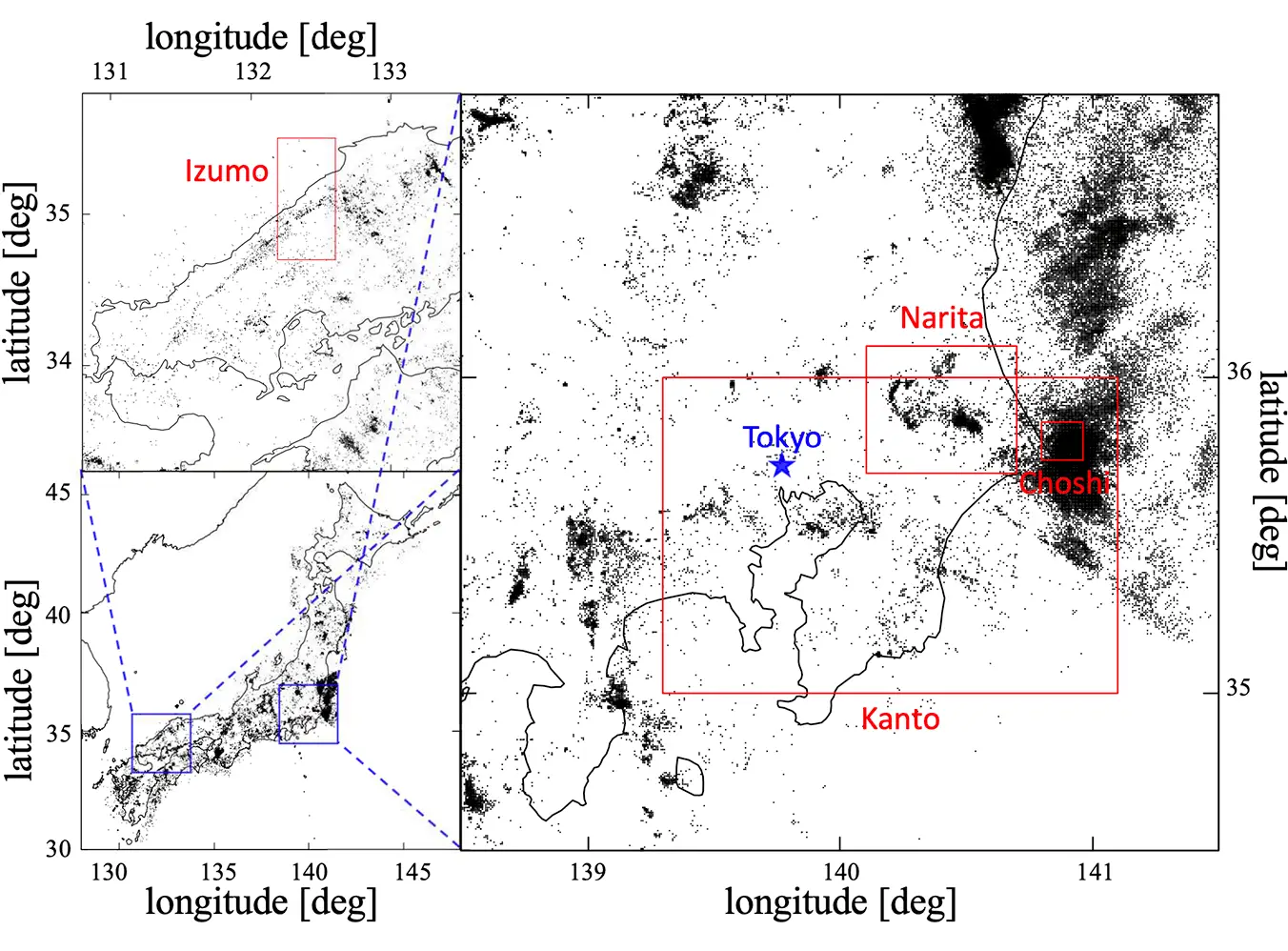
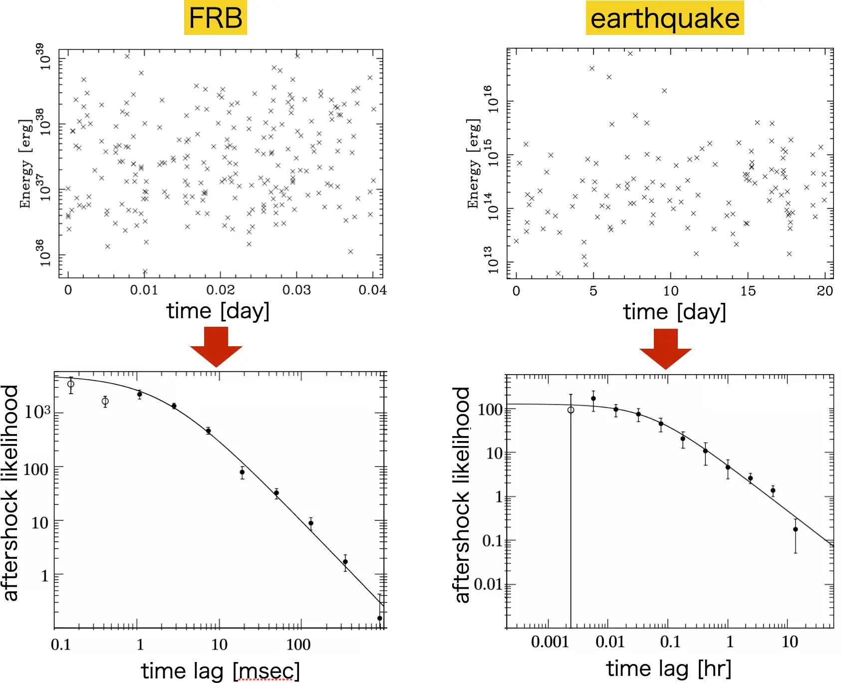
So far, statistical analysis of FRBs has focused on the distribution of wait times between two successive bursts. However, Totani and co-author Yuya Tsuzuki, a graduate student in the same department, point out that calculating only the wait-time distribution does not take into account correlations that might exist across other bursts. So the team decided to calculate correlation across two-dimensional space, analyzing the time and emission energy of nearly 7,000 bursts from three different repeater FRB sources. They then applied the same method to examine the time-energy correlation of earthquakes (using data from Japan) and solar flares (using records from the Hinode international mission to study the sun), and compared the results of all three phenomena.
Totani and Tsuzuki were surprised that, in contrast to other studies, their analysis showed a striking similarity between FRBs and earthquake data, but a distinct difference between FRBs and solar flares.
Totani explained: “The results show notable similarities between FRBs and earthquakes in the following ways: First, the probability of an aftershock occurring for a single event is 10-50%; second, the aftershock occurrence rate decreases with time, as a power of time; third, the aftershock rate is always constant even if the FRB-earthquake activity (mean rate) changes significantly; and fourth, there is no correlation between the energies of the main shock and its aftershock.”
This strongly suggests the existence of a solid crust on the surface of neutron stars, and that starquakes suddenly occurring on these crusts release huge amounts of energy which we see as FRBs. The team intends to continue analyzing new data on FRBs, to verify that the similarities they have found are universal.
“By studying starquakes on distant ultradense stars, which are completely different environments from Earth, we may gain new insights into earthquakes,” said Totani. “The interior of a neutron star is the densest place in the universe, comparable to that of the interior of an atomic nucleus. Starquakes in neutron stars have opened up the possibility of gaining new insights into very high-density matter and the fundamental laws of nuclear physics.”
Reference: “Fast radio bursts trigger aftershocks resembling earthquakes, but not solar flares” by Tomonori Totani and Yuya Tsuzuki, 11 October 2023, Monthly Notices of the Royal Astronomical Society.
DOI: 10.1093/mnras/stad2532