

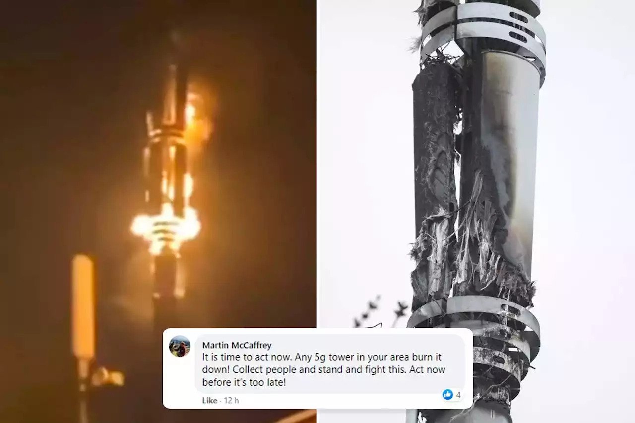
We all know that the Coronavirus is causing havoc across the globe. It started off in Wuhan, China, and has spread across the world. Recently a bunch of crazy conspiracy theorists have thought to have linked the virus to 5G, and have started destroying masts wherever they see them.
Destruction of 5G masts in UK
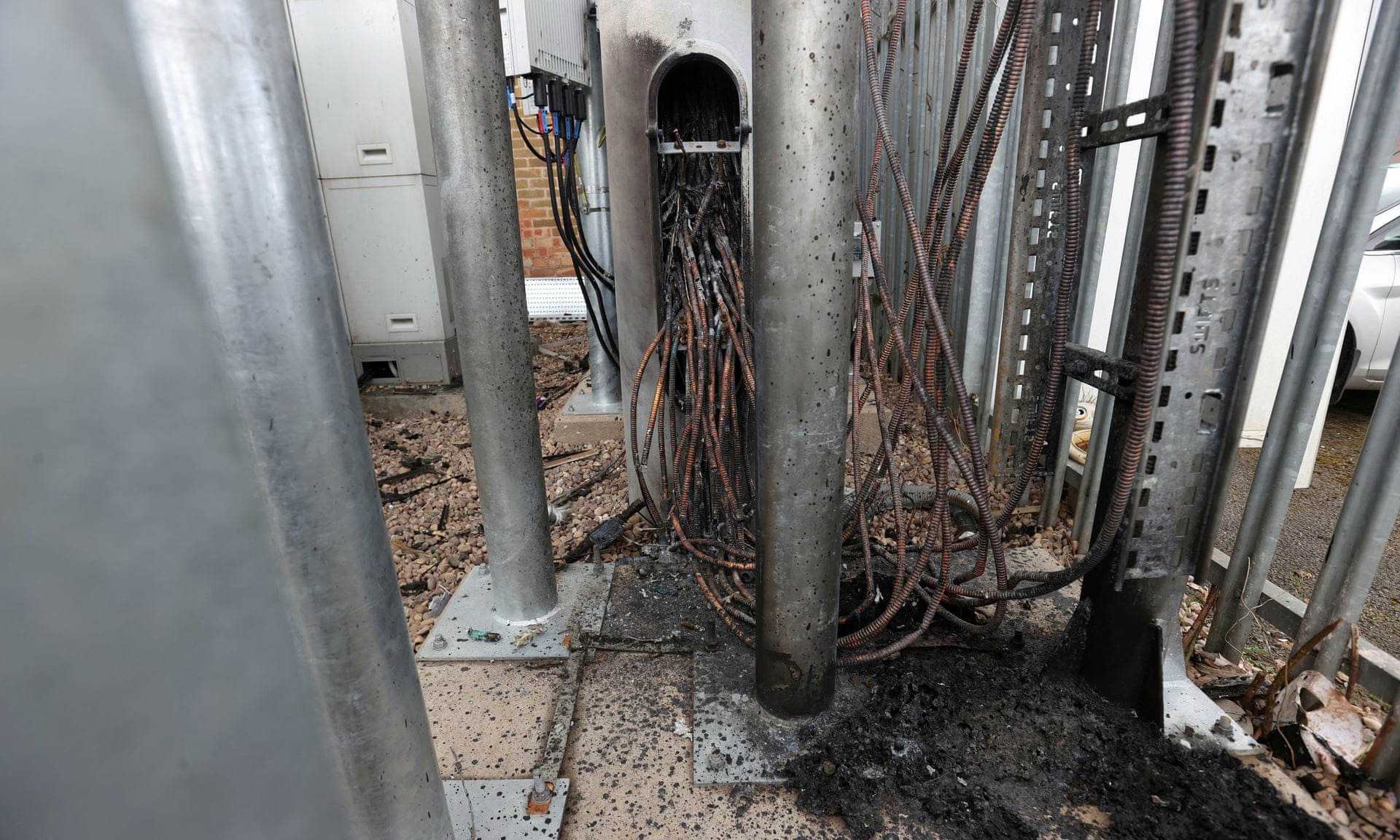
At least 20 5G masts across the UK have been torched or vandalised across the UK since Thursday the 2nd of April 2020. According to govt sources, they are linking the Coronavirus to the installation of 5G networks across the country. Multiple attacks on 5G masts have been noticed in Liverpool and the West Midlands. Due to the slow rollout of 5G, many 3G and 4G masts too have been damaged. There seems to be some hope that the attacks have reduced for now. Network operators are concerned for the safety of their staffs as attackers have confronted the network engineers, and even filming the encounters. There have been around 30 incidents in the past week.
Educating the masses
Social media platforms will soon partner with the government in order to educate the masses and do what they can to stop the baseless spread of 5G being linked to the virus. They can hardly do much with the spread of misinformation on WhatsApp. There have been repeated assurances by international radiation watchdogs claiming that 5G is safe, but the baseless theories have taken over, leaving the truth in the dust. Recently, the rapid explosion of claims has caught the eyes of celebrities, and the Industry wasn’t aware of it. There have been many long-running Facebook groups that tried to increase the spread of misinformation by opposing the rollout of 5G, by further encouraging vandalism of the 5G network. Facebook has tried its best to delete as many of those groups as they can.
After the arson attack of masts in Birmingham, the attacks grew rapidly. Vodafone confirmed that six sites were targeted over the weekend, and other networks suggested they had seen similar numbers.MobileUK, the industry group that unites the UK’s four main mobile networks, published an open letter to customers asking for help to stop the vandalism.
“We have experienced cases of vandals setting fire to mobile masts, disrupting critical infrastructure and spreading false information suggesting a connection between 5G and the Covid-19 pandemic,” the open letter says. “There is no scientific evidence of any link between 5G and coronavirus.” Fact.
“Please help us to make this stop. If you witness abuse of our key workers please report it. If you see misinformation, please call it out. Your help will make a real difference.”
What do you think? Would you help stop the fake theories of 5G being responsible for the spread of Coronavirus in the UK? Do let us know in the comments below.
Source: The Guardian