HTC has been introducing many great and new stuffs in the tech industry like the HTC Eye Camera and more. But, what about a Attachable Camera for Smartphones? Better Pictures from your Phone? Better Selfies? Obviously Yes! So… Here we bring an hot concept of such a device which is yet unofficial but we do wish it to be actually Official! 😀 Introducing HTC MiniCam!
If you’ve been keeping up with the latest rumors surrounding the future release of the Motorola Moto X 2016, you probably know that one of its versions will accept back plate modules. Why not envision something similar for HTC?
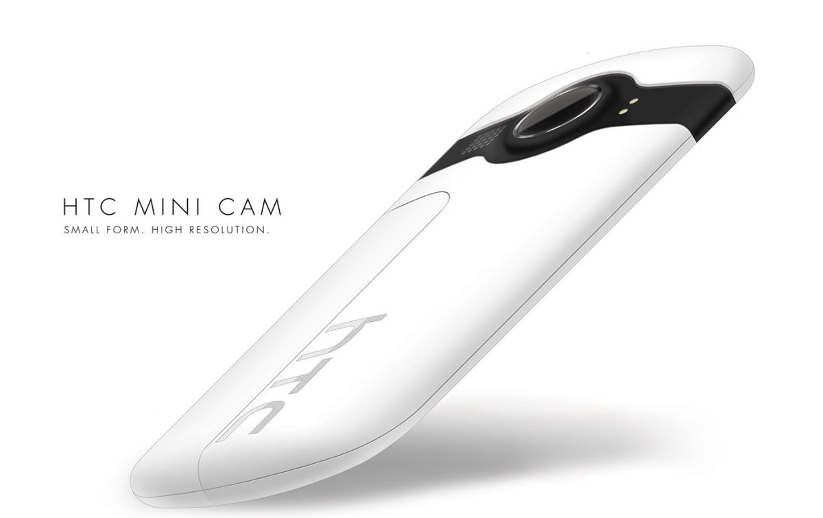
Designer William Turner from Miami has created the HTC MiniCam, an attachable and adaptable camera module for the HTC phones and probably for more devices. The idea was to create a pocked sized camera, that attaches to a smartphone and lets the user take higher resolution pictures. The attachment also brings a front facing flash and extra features, possibly laser focus and depth cameras.
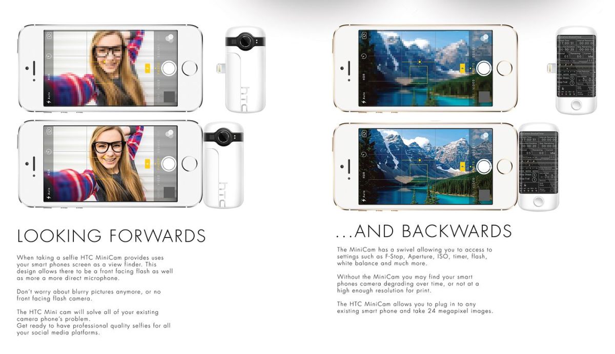
The camera module can be swiveled in front or behind the phone, in order to amplify its features. The module has a touchscreen at the back, with manual and auto settings shown. It can arrive with a Lightning or microUSB port and it measures about one fourth of an iPhone, so it’s not very bulk and doesn’t add that much to the phone’s length.
24 megapixel shots are promised to be made by using the HTC MiniCam. Which sounds pretty amazing and ofcource, who doesn’t wants such a powerful shooter for your smartphones especially when your device has like 5MP or 8MP cameras and you struggle for shots! Great Concept So Far!
Hey! You there, let us know whats up on your mind about this and shoot us with a comment down below and lets begin a healthy Conversation/Discussion about this product! Waiting…