Dutch retailer Centralpoint is offering tray versions of AMD’s 65w top-end Ryzen 4000 Renoir desktop APUs. While these price might look a lot, it would be safe to assume that the price in the US would be way lower than what it currently is at Centralpoint. The evidence of the launch of the new APU lineup was uncovered by Twitter user momomo_us, who caught sight of the prices on the Centralpoint site.
Ryzen 4000 Renoir Pro processors list
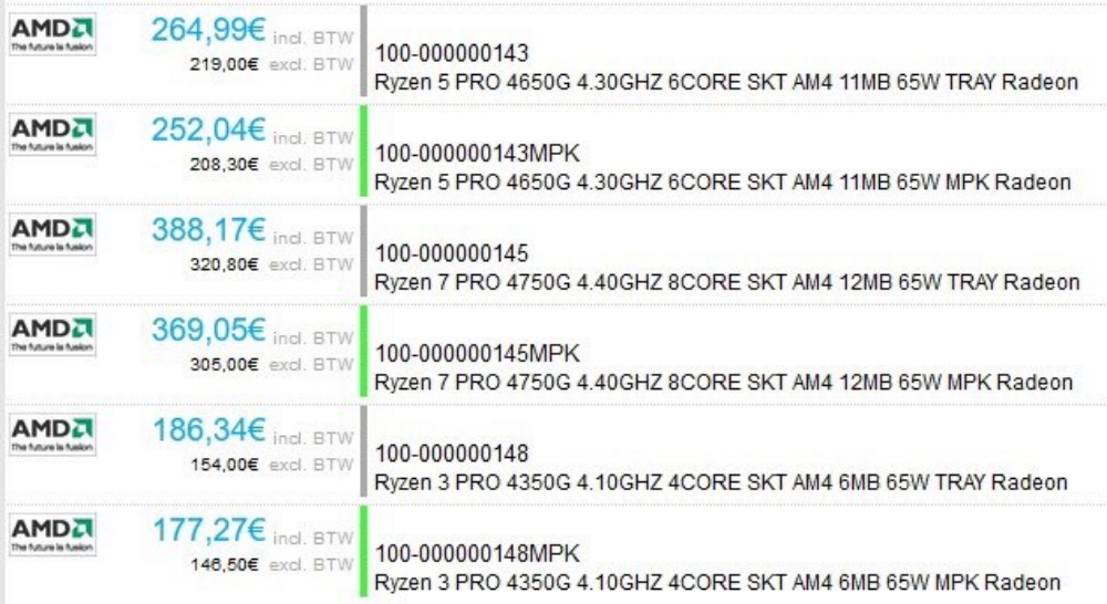
The Ryzen 4000 APUs were having a bunch of leaks for the past few months, and as we can see from this listing, the launch is just around the corner. So it’s evident that AMD will be using a new naming scheme to differentiate between the regular desktop and professional users. The Dutch retailer has listed 3 variants of the Pro series. They are Ryzen 7 Pro 4750G, Ryzen 5 Pro 4650G and Ryzen 3 Pro 4350G. There are 2 variants for each processor. The first is the tray variant, and the second is the boxed variant. Tray processors are those handed to OEMs(Original Equipment Manufacturers) who manufacture pre-assembled desktops and laptops. Warranty replacements and technical support for tray processors are provided directly by these OEMs. CPU companies don’t provide direct warranty support.
AMD Ryzen 7 Pro 4750G
This is the flagship of the Renoir PRO desktop APUs. It uses TSMC’s 7nm process on the Zen2 architecture as well as the 7nm process on the Vega GPU too. It uses 8 cores and 16 threads. The base clock is set at 3.6GHz while boost is at 4.4GHz. The combined cache is 12MB (4MB L2 + 8MB L3). TDP is 65w and the GPU is Vega 8 with 512 stream processors clocked at 2.1GHz. There are currently placeholder prices on the site, and they could be updated after the launch. As seen in the above pic, the boxed and tray versions are 388.17 and 369.05 Euros (VAT included) respectively.
AMD Ryzen 5 Pro 4650G
The midrange AMD Ryzen 5 Pro 4650G is a 6 core and 12 thread APU. It has a base clock of 3.70 GHz and a boost clock of 4.30 GHz. The combined cache here is 11 MB (3MB L2 + 8MB L3) with a TDP of 65W. GPU features the Vega 7 chip with 448 stream processors clocked at 1.9 GHz. The boxed variant of the Ryzen 5 Pro 4650G costs 252.04 Euros, while the tray variant comes to 264.99 Euros.
AMD Ryzen 3 Pro 4350G
Last but not the least, the entry level AMD Ryzen 3 Pro 4350G Renoir PRO Desktop CPU is a 4 cores 8 threaded APU. The APU features a base clock of 3.80 GHz and a boost clock of 4.10 GHz. There’s 6 MB of cache & the CPU features a TDP of 65W. The graphics side includes the Vega 6 chip with 384 SPs clocked at 1700 MHz. The boxed variant of the Ryzen 3 Pro 4350G costs 177.27 Euros while the tray variant costs 186.34 Euros.
Currently, there’s no release date announced, but the fact that these APUs were listed in a Dutch retailer’s shop proves that the launch is right around the corner, which means maybe a month or two.